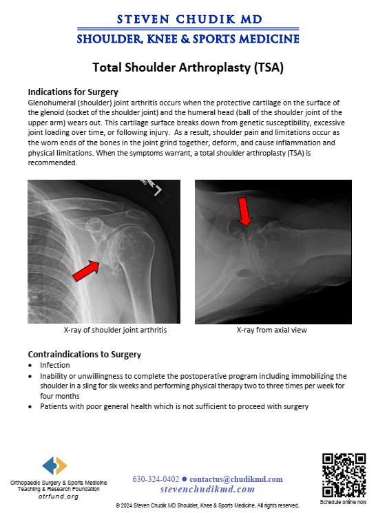
Total Shoulder Arthroplasty (TSA)

RESEARCH A Novel Anatomic Portal for Shoulder Replacement

Articular cartilage lesions and arthritis involving the glenohumeral joint can be difficult to treat. Conventional methods include arthroscopic debridement, abrasionplasty, microfracture, cartilage restoration procedures, resurfacing and replacement. Restoration, resurfacing, and replacement methods achieve limited access to the glenohumeral joint following subscapularis transection and dislocation. Subscapularis transection delays rehabilitation often resulting in decreased range of motion and permanent weakness. Dr. Steven Chudik developed novel techniques and instruments to access the articular surface without subscapularis transection or dislocation of the glenohumeral joint, allowing for partial and complete osteochondral transplantation or artificial replacement of the articular surfaces.

We propose that utilizing a novel transhumeral portal through the neck of the humerus can decrease surgical morbidity and improve our ability to restore the normal anatomic relationships of the glenohumeral joint. This portal not only is less disruptive to the surrounding structures, but also accounts for patient variability when replacing the humeral surface by referencing from the center of the humeral head and neck. This differs from conventional methods that use the intrameduallary canal, which has an inconsistent relationship to the humeral surface.

This study investigated the safety and efficacy of using a novel transhumeral portal to access and prepare the articular surfaces of the glenohumeral joint. The transhumeral portal demonstrated reliable perpendicular access to the glenohumeral joint surface without surgical dislocation, humeral head resection, nor injury to the proximate important anatomic structures, particularly the axillary nerve and rotator cuff. Although there still remains limitations with regards to judging the center-center point on the humeral head using arthroscopic guidance alone, this novel transhumeral portal possesses great potential for decreased surgical morbidity, more anatomic replacement of the glenohumeral surfaces, immediate active range-of-motion and strengthening, and better functional outcomes for patients than conventional open approaches.

RESEARCH The Glenoid Vault: Anatomic Cadaver Study Exploring Alternative Solutions for Glenoid Implant Survival in Total Shoulder Arthroplasty (TSA)

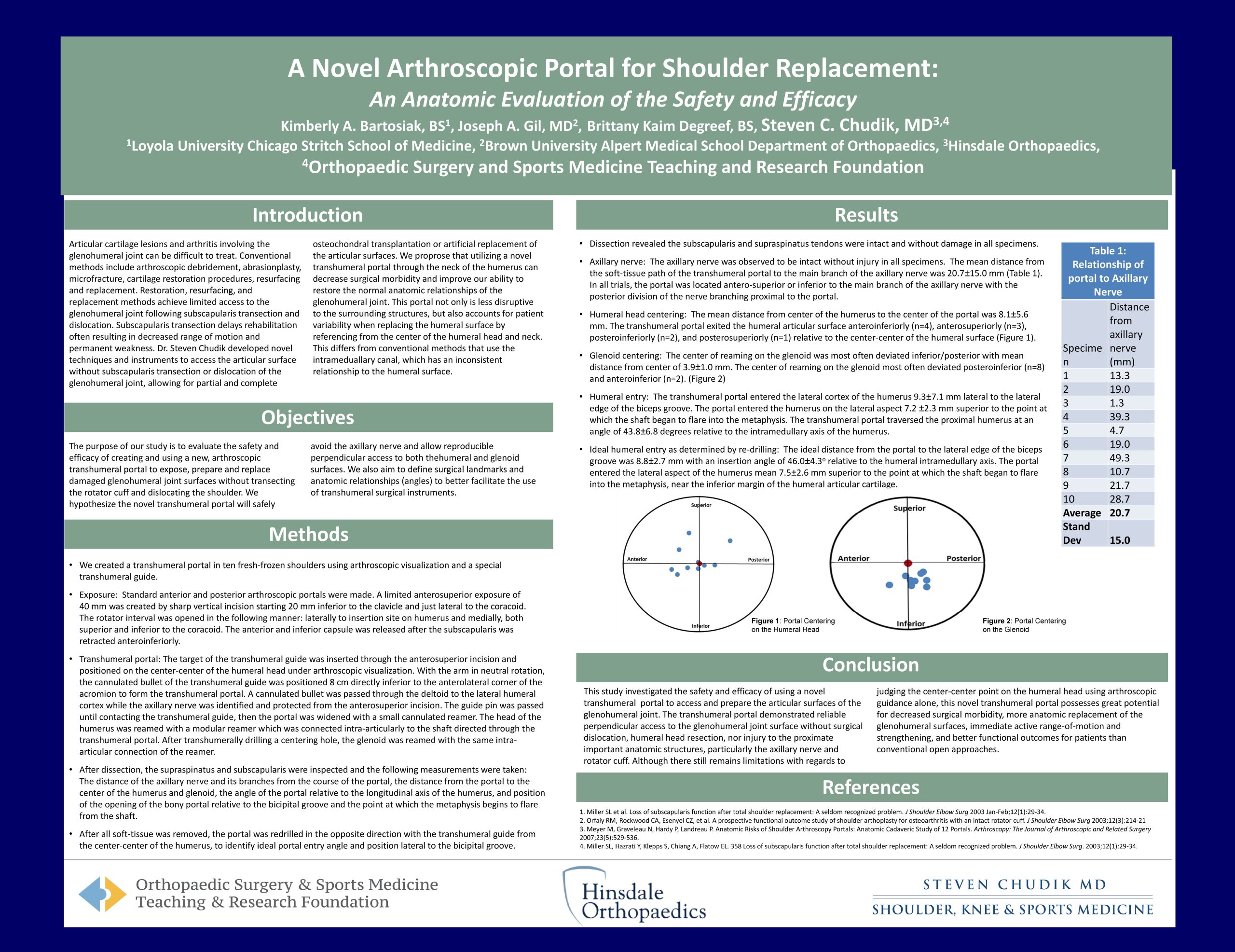
Total shoulder arthroplasty is a common surgical procedure indicated for arthritis of the glenohumeral joint. Although this surgical procedure is highly successful in restoring the function of the glenohumeral joint and relieving pain associated with glenohumeral degeneration, it is not free of post-operative complications. Prosthetic loosening accounts for 39% of total shoulder arthroplasty complications, with glenoid component loosening being responsible for 32% of all complications. Glenoid component loosening is one of the most common causes of total shoulder arthroplasty failure, and is associated with symptoms such as pain, stiffness, material failure, wear and joint instability. Further, revision surgery is indicated in most cases of glenoid component loosening. The common problem of glenoid component loosening is one that has not been fully resolved since the development and application – thus, there is a clear and enduring need to further explore the glenoid’s morphology and discover alternative methods for stabilization of the glenoid component in total shoulder arthroplasty.

Through examination of the anatomy of the glenoid vault as it pertains to glenoid component implantation, there is potential to better understand the cause of glenoid component loosening and craft new approaches to ensure the stability and longevity of the implant. While many anatomic studies have been conducted to quantify the superficial features of the glenoid, fewer have investigated its internal spatial composition in both a qualitative and quantitative manner – thus, digitization of the glenoid and its surrounding scapular structures using our study’s protocol has the potential to yield novel information about its overall morphology and capacity for modification.

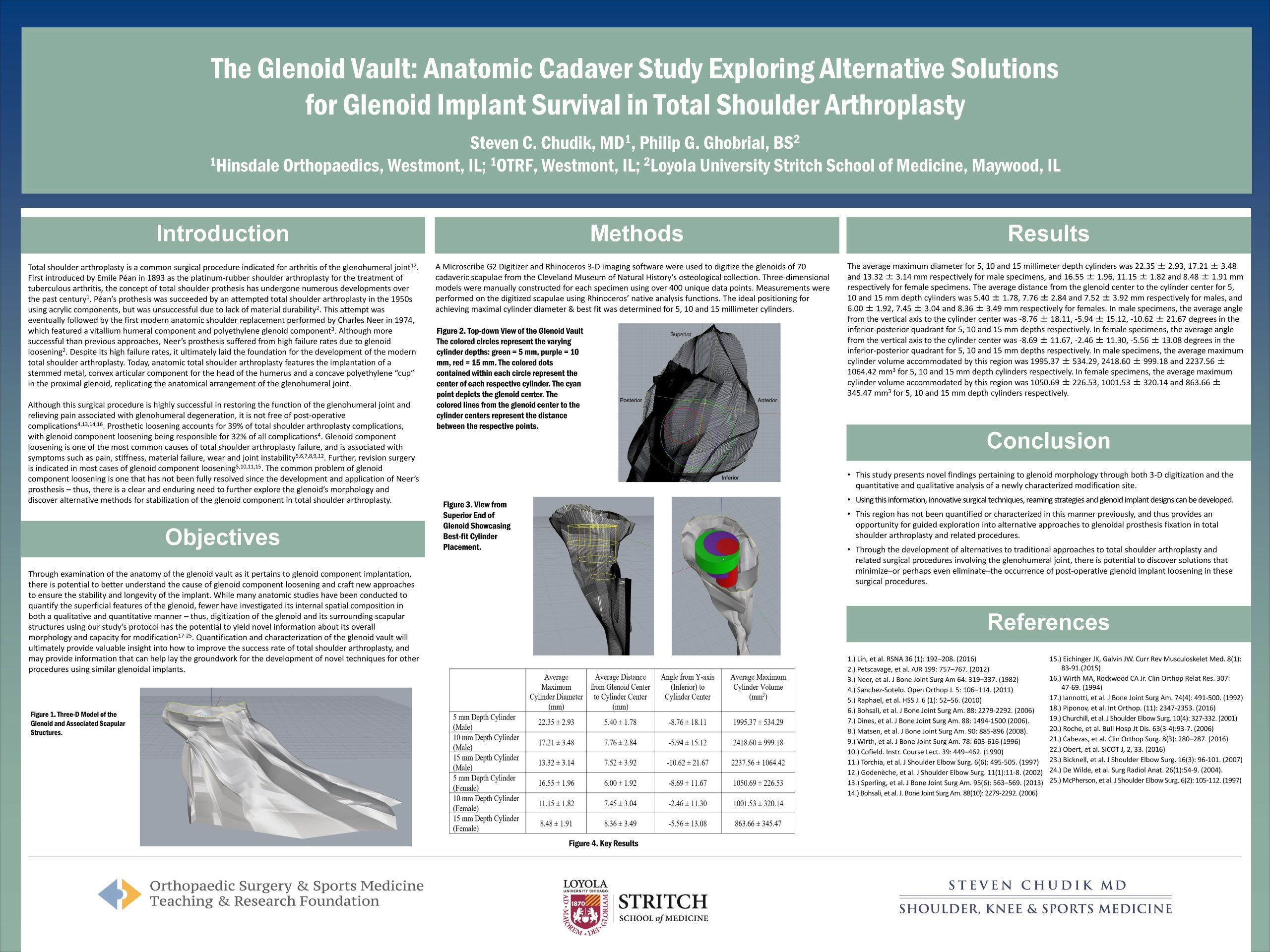
Content provided by Dr. Chudik not to be used for diagnosis and treatment. You can receive a proper evaluation and diagnosis by making an appointment with Dr. Chudik

Western Springs Office

Wed: 7:30 a.m. to 5:30 p.m.

4700 Gilbert Avenue, Suite 51
Western Springs, Illinois 60558
Phone: 630-324-0402
Fax: 630-920-2382

right-footer-logo

Steven Chudik MD Shoulder and Knee Injury Clinic.

© 2023 © 2019 Copyright Steven Chudik MD, All Rights Reserved.

Privacy Policy and Disclaimer
WordPress Video Lightbox